Proteins are the cell's workhorses, and proteomics reveals their dynamic activity. The genetic code is just the beginning. Post-translational modifications (PTMs) introduce a layer of complexity to the proteome, enabling proteins to adopt diverse structures and functions. Changes in protein levels can serve as early indicators of disease, making proteomics a powerful diagnostic tool. By analyzing protein expression, researchers can unlock the secrets of cellular response to stimuli like stress, disease, and treatments. When combined with genomics and transcriptomics, proteomics provides a comprehensive view of biological systems. This multi-omics approach accelerates the discovery of key molecular players and pathways involved in complex diseases, paving the way for novel biomarkers and therapeutic targets.
Read more about Spatial Biology here

Spatial proteomics has historically lagged behind spatial transcriptomics in terms of multiplexing and throughput due to the inherent complexities of proteins, such as their diverse shapes and sizes, intricate three-dimensional structures, and the inability to amplify them like DNA. However, recent breakthroughs have significantly advanced spatial proteomics. These include the development of antibody-based imaging technologies like Imaging Mass Cytometry (IMC-Cytof) and Multiplexed Ion Beam Imaging by Time-of-Flight (MIBI-TOF), enabling the simultaneous detection of multiple proteins within their spatial context. Furthermore, the adaptation of highly sensitive Liquid Chromatography-Mass Spectrometry (LC-MS) -based proteomics to spatial biology has further expanded the capabilities of this field. Spatial CITE-seq (spatial co-indexing of transcriptomes and epitopes for multi-omics mapping by NGS), with a wide range of applications in biomedical research has the potential to detect > 200 protein markers in situ along with transcriptome via (Deterministic Barcoding) DBiT-seq technology, using a cocktail of antibody-derived DNA tags (ADTs).
A novel spatial proteomics technology called DISCO-MS (three-dimensional imaging of solvent-cleared organs profiled by mass spectrometry) in optically cleared 3D intact specimens enables profiling of complex tissues to investigate cellular functions in physiological and pathological state.
DISCO-MS integrates advanced 3D imaging, AI-powered analysis, and robotic tissue extraction to identify and isolate targeted tissue regions for subsequent mass spectrometry analysis. While the current resolution is not at the single-cell level, DISCO-MS provides a powerful platform for investigating tissue heterogeneity and disease mechanisms by enabling comprehensive spatial proteome analysis of whole organs.
Earlier breakthroughs in spatial proteomics did not have single-cell (SC) resolution capabilities. Most sc technologies focused on the nuclei acids (DNA and RNA) A new era of single-cell proteomics has emerged, thanks to breakthroughs in sample preparation, chromatographic separations, and mass spectrometry. This enables researchers to go deeper into the proteomic landscape of individual cells.
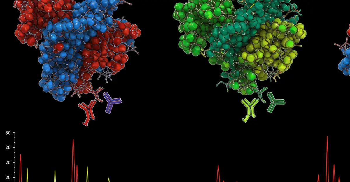
There are two main approaches for SC proteomics. Antibody-based proteomics leverages flow cytometry to measure proteins in single cells using fluorescent-tagged antibodies. Mass cytometry, an advanced technique, employs metal-tagged antibodies for simultaneous detection of multiple proteins via mass spectrometry. This method offers higher multiplexing capability compared to flow cytometry. Additionally, imaging mass cytometry allows for spatial protein mapping in tissue sections. Another approach is Mass spectrometry (MS) , a powerful tool for analyzing the entire proteome by measuring the mass-to-charge ratio of molecules. While traditionally used for bulk analysis, recent advancements in sample preparation techniques have enabled single-cell proteomics. However, limitations in throughput remain. New mass spectrometry methods, including SCoPE2 (Single Cell ProtEomics by Mass Spectrometry), overcome these challenges by quantifying many proteins per cell with high specificity. Developed by researchers at Northeastern University Boston, SCoPE2 uses isobaric carrier technology to improve peptide identification. It involves isolating single cells, lysing them, labeling peptides with isobaric tags, and analyzing them using mass spectrometry. SCoPE2 is cost effective, scalable, efficient and versatile.
Scientists at The Max Delbrück Center for Molecular Medicine revolutionized SC Proteomics with the development of Deep Visual Proteomics (DVP), a new concept that combines fluorescence or brightfield imaging based single-cell phenotyping with unbiased MS (Mass Spectrometry)-based proteomics for global proteome profiling with cell type and spatial resolution.
DVP involves laser microdissection to isolate specific regions from complex samples. Subsequently, individual cells within these dissected regions are analyzed using mass spectrometry, while meticulously preserving their spatial origin. This unique approach offers a significant advantage over antibody-based methods, as it is not constrained by the availability of specific antibodies. Consequently, DVP enables substantially broader proteome coverage, providing a more comprehensive understanding of protein distribution and function within the spatial context of the tissue. A workflow developed especially for DVP, easy-to-use, robust and scalable was applied to study the cell type resolved proteome of B and T lymphocytes with use of as little as 100 phenotype-matched cells from archival tissue material, while preserving detailed cell type and spatial information.
Spatial proteomics, in conjunction with the Human Protein Atlas (HPA), offers a powerful approach to understanding the spatial organization of the human proteome. HPA initiative utilizes advanced omics technologies to provide open-access data on protein expression, localization, and interactions. By integrating information from various resources, researchers can explore the human proteome, identify disease biomarkers, and uncover novel therapeutic targets. The Atlas has already made significant contributions to the field of human biology and is recognized as a vital resource for the global scientific community.
Recognized for its groundbreaking contributions, Spatial Proteomics was named Method of the Year by Nature Methods in December 2024.